Advance Dental Center © Copyright 2015 | All Rights Reserved|Sitemap
X-rays and CT Scans
 Diagnostic imaging techniques help narrow the causes of an injury or illness and ensure that the diagnosis is accurate. These techniques include X-rays and computed tomography (CT) scans.
These imaging tools let your doctor “see” inside your body to get a “picture” of your bones, organs, muscles, tendons, nerves, and cartilage. This is a way the doctor can determine if there are any abnormalities.
X-rays
X-rays (radiographs) are the most common and widely available diagnostic imaging technique. Even if you also need more sophisticated tests, you will probably get an X-ray first.
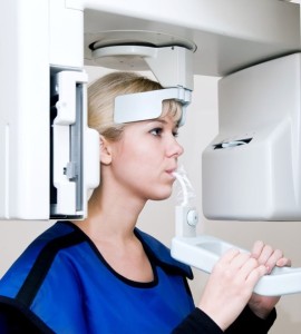
The part of your body being pictured is positioned between the X-ray machine and photographic film. You have to hold still while the machine briefly sends electromagnetic waves (radiation) through your body, exposing the film to reflect your internal structure. The level of radiation exposure from X-rays is not harmful, but your doctor will take special precautions if you are pregnant.
Bones, tumors and other dense matter appear white or light because they absorb the radiation. Less dense soft tissues and breaks in bone let radiation pass through, making these parts look darker on the X-ray film. Sometimes, to make certain organs stand out in the picture, you are asked given barium sulfate or a dye.
You will probably be X-rayed from several angles. If you have a fracture in one limb, your doctor may want a comparison X-ray of your uninjured limb. Your X-ray session will probably be finished in about 10 minutes. The images are ready quickly.
X-rays may not show as much detail as an image produced using newer, more powerful techniques.